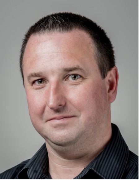
Fiche de LALANDE Alain

| Adresse 1: | Laboratoire ICMUB, Faculté de Médecine, 7 Bld Jeanne d’Arc, BP 87900, 21079 ,Dijon, cedex, FRANCE |
| Adresse 2 : | Pôle Imagerie, Hôpital d’Enfant - CHU de Dijon 1 Bld Jeanne d’Arc, BP 77908, 21079 Dijon Cedex, FRANCE |
| Tél : | +33 3 80 39 33 91 |
| Email : | alain.lalande@ube.fr |
| Equipe : | IFTIM |
| Fonction : | Directeur d'unité adjoint Maître de conférences |
| ORCID ID : | 0000-0002-7970-366X |
- Carrière
- Projets
- Publications
- Enseignements
Mai 2012 : « Habilitation à Diriger les Recherches » en Sciences, Dijon (France)
Sept 2000 : Maitre de conférences, Université de Bourgogne, Dijon (France)
April 1999 : Doctorat, Université de Bourgogne, Dijon (France)
1995-1997,1999 : ATER en Biophysique, Université de Bourgogne, Dijon (France)
1998 : Ingénieur en Informatique (Service Militaire), Saint Mandé (France)
Juin 1995: Master en Informatique en médecine, Paris (France).
- ADEPT ( ) . Financement FEDER (Programme FEDER-FSE+ Bourgogne Franche-Comté et Massif du Jura 2021-2027).
- BosomShield ( https://bosomshield.eu/ ) . Financement européen de type Marie Sklodowska-Curie Doctoral Networks Actions (HORIZON-MSCA-2021-DN-01-01)
- Co-organisateur du challenge ACDC lors de la conférence MICCAI 2017 et responsable de la base de données associé.
- Organisateur du challenge EMIDEC lors de la conférence MICCAI 2020 et responsable de la base de données associé.
Liste complète de publications
- D.M. Marin-Castrillon, A. Lalande, S. Leclerc, K. Ambarki, M.-C. Morgant, A. Cochet, S. Lin, O. Bouchot, A. Boucher, B. Presles. 4D segmentation of the thoracic aorta from 4D flow MRI using deep learning. Magnetic Resonance Imaging. 2023;99:20-25
- A. Lalande, Z. Chen, T. Pommier, T. Decourselle, A. Qayyum, M. Salomon, et al. Deep learning methods for automatic evaluation of delayed enhancement-MRI. The results of the EMIDEC challenge. Medical Image Analysis, 2022 ; 79 : 102428.
- Y. Skandarani, A. Lalande, J. Afilalo, P.M. Jodoin. Generative adversarial networks in cardiology. Canadian Journal of Cardiology 38 (2), 196-203.
- K.B. Girum, G. Créhange, A. Lalande. Learning with Context Feedback Loop for Robust Medical Image Segmentation. IEEE Trans Med Imaging, 2021; 40(6):1542-1554. doi: 10.1109/TMI.2021.3060497.
- N. Painchaud, Y. Skandarani, T. Judge, O. Bernard, A. Lalande, P.M. Jodoin . Cardiac Segmentation With Strong Anatomical Guarantees. IEEE Trans Med Imaging. 2020 Nov;39(11):3703-3713.
- R. Hussain, A. Lalande, C. Guigou, A. Bozorg-Grayeli. Contribution of Augmented Reality to Minimally Invasive Computer-Assisted Cranial Base Surgery. IEEE J Biomed Health Inform. 2020 Jul;24(7):2093-2106.
- R. Hussain, A. Lalande, R. Marroquin, C. Guigou, A. Bozorg Grayeli. Video-based augmented reality combining CT-scan and instrument position data to microscope view in middle ear surgery. Sci Rep. 2020 Apr 21;10(1):6767.
- J.Z. Bojorquez, P.M. Jodoin, S. Bricq, P.M. Walker, A. Lalande. Automatic classification of tissues on pelvic MRI based on relaxation times and support vector machine. Plos One, 2019; 14(2):e0211944
- O. Bernard, A. Lalande, C. Zotti, F. Cervenansky, X. Yang, PA Heng, I Cetin, et al. Deep Learning Techniques for Automatic MRI Cardiac Multi-Structures Segmentation and Diagnosis: Is the Problem Solved? IEEE Trans Med Imaging, 2018; 37(11), 2514-2525.
- J.Z. Bojorquez, S. Bricq, C. Acquitter, F. Brunotte, P.M. Walker, A. Lalande. What are normal relaxation times of tissues at 3Tesla? Magn Reson Imaging, 2017; 35:69-80.
- L. Bal-Theoleyre, A. Lalande, F. Kober, R. Giorgi, F. Collart, P. Piquet, G. Habib, J.F. Avierinos, M. Bernard, M. Guye, A. Jacquier. Aortic Function’s Adaptation in Response to Exercise-Induced Stress Assessing by 1.5T MRI: A Pilot Study in Healthy Volunteers. PLoS One. 2016 Jun 16;11(6):e0157704.
- P.-M. Jodoin, F. Pinheiro, A. Oudot, A. Lalande. Left-ventricle segmentation of SPECT images of rats. IEEE Trans Biomed Eng. 2015; 62(9): 2260-2268.
- Y. Wang, D. Joanic, P. Delassus, A. Lalande, P. Juillon, J.-F. Fontaine. Comparison of the strain field of abdominal aortic aneurysm measured by magnetic resonance imaging and stereovision: a feasibility study for prediction of the risk of rupture of aortic abdominal aneurysm. J Biomech. 2015; 48(6): 1158-1164.
- M. Xavier, A. Lalande, P.M. Walker, F. Brunotte, L. Legrand. An adapted optical flow algorithm for robust quantification of cardiac wall motion from standard cine-MR examinations. IEEE Trans Inf Technol Biomed. 2012; 16(5):859-868.
- A. Cochet, A. Lalande, L. Lorgis, M. Zeller, J.C. Beer, P.M. Walker, C. Touzery, J.E. Wolf, Y. Cottin, F. Brunotte. Prognostic Value of Microvascular Damage Determined by Cardiac Magnetic Resonance in Non ST-Segment Elevation Myocardial Infarction: Comparison Between First-Pass and Late Gadolinium-Enhanced Images. Invest Radiol, 2010; 45(11): 725-732.
- A. Lalande, P. Khau Van Kien, P.M. Walker, L. Zhu, L. Legrand, M. Claustres, X. Jeunemaître, F. Brunotte, J.-E. Wolf. Compliance and pulse wave velocity assessed by MRI detect early aortic impairment in young patients with mutation of the smooth muscle myosin heavy chain. J Magn Reson Imaging, 2008; 28 (5): 1180-1187.
- L. Zhu L, R. Vranckx R, P. Khau Van Kien, A. Lalande , N. Boisset, F. Mathieu, M. Wegman, L. Glancy, J.M. Gasc, F. Brunotte, P. Bruneval, J.E. Wolf, J.B. Michel, X. Jeunemaitre. Mutations in myosin heavy chain 11 cause a syndrome associating thoracic aortic aneurysm/aortic dissection and patent ductus arteriosus. Nature Genetics, 2006; 38(3):343-349.
Cursus des études médicales
PACES. Enseignement en biophysique
P2. Enseignement en biophysique
UE Imagerie fonctionnelle et moléculaire. Enseignement en imagerie médicale
Odontologie (DFGSO3). Enseignement en imagerie par rayons X
DU IA Santé. Responsable du module « Imagerie médicale et IA ». Enseignement en imagerie médicale et intelligence artificielle
Masters
Master I MAIA (Master Erasmus Mundus in Medical Imaging and Applications). Responsable du module Sensors and digitization. Enseignement en imagerie médicale.
Master II Health AI. Responsable du module Sensors and digitization. Enseignement en imagerie médicale.
Master II TSI (Traitement du Signal et des Images). Responsable du module « De l’image aux applications médicales ». Enseignement en imagerie médicale.
Master II Innovative Drugs. Enseignement en traitement d’images médicales
Directeur de thèse
You ZHOU. Détermination automatique à partir d’IRM de paramètres biomécaniques comme facteurs de risque dans le cadre d’anévrisme de l’aorte thoracique
Co-directeur de thèse
Abulrahman ALBLOWI. Les caractéristiques biomécaniques et histologique des différents patchs péricardiques bovins conservés dans une solution de glutaraldéhyde (co-tutelle avec l’Université de Lorraine)
Tewele W. TAREKE. Analyse d’imagerie multi-modale : corrélation des biomarqueurs entre imagerie radiologique et histopathologique